Registration (aka spatial normalization)
A goal common to many neuroimaging studies is to learn something about how patterns of brain activity generalize from the volunteers tested to a broader population. In the case of MRI, this generally involves registering a group of participants’ brains into a common space. If the patterns of neural activation have a systematic relationship to macroanatomical structure (see below), then lining up these structures across volunteers allows us to make inferences about the level of group activity.
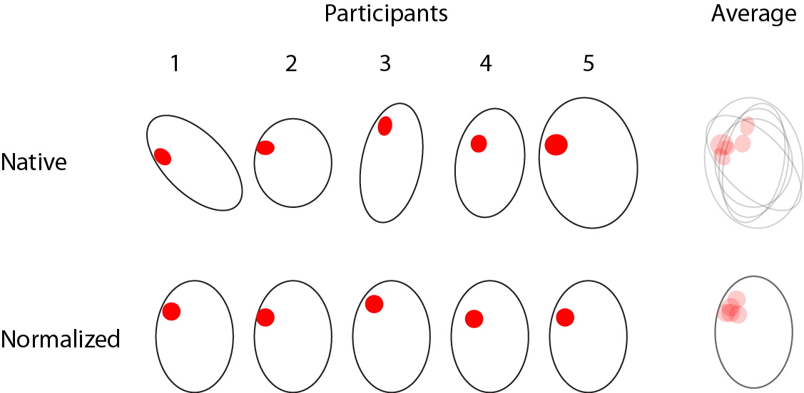
Schematic illustration of 5 participants showing “activation” in a similar location in the brain. Without registering these brains to a common space, these images cannot be easily compared (top). When they are (bottom), the overlap is evident.
If the common space is also a standard space, such as MNI space (see below), then registration also facilitates the sharing and interpretation of data across studies. This could involve either the reporting of peak coordinates in MNI space (millimeters along X, Y, and Z axes) or the comparison of whole-brain parameter estimates.
MNI space vs. “Talairach” space
The Talairach and Tournoux atlas ([TalairachTournoux1988]) describes a stereotaxic reference frame for the human brain (see “Talairach” space). This reference frame, generally speaking, can be applied to any brain. In that sense, any number of “spaces” might be referred to as “Talairach space”, because they follow the same general stereotactic convention. However, in the Talairach and Tournoux atlas, the brain is that of a single adult, and “Talairach space” is best used when referring to the space described by that brain (see also [Devlin2007])
The Montreal Neurological Institute (MNI) developed several templates based on averaging a large number of adult brains. Because these templates are based on a group average, they better represent a “standard” brain than a single brain will. The templates included with SPM (and most other neuroimaging analysis packages) are in MNI space. These templates have been adopted by the International Consortium for Brain Mapping (ICBM; http://www.loni.ucla.edu/ICBM/) as a standard, and so sometimes are referred to as being in ICBM space.
Note
Templates are commonly referred to by the number of brains that went into their creation; thus, ICBM152 space refers to the ICBM template created using 152 brains (which, in the case of ICBM152, were spatially normalized to an earlier template, MNI305).
Correspondence between macroanatomical and cytoarchitectural characteristics
Most of the time, what we see visually in an MRI image—and the differences in image intensity values that are used in image registration—is attributed to macroanatomy, or patterns of cortical folding. However, it is important to remember that although on an MRI scan (or on a real brain) gray matter has a consistent look throughout the brain, it is in fact made up of a number of different types of cells. The different types of cells, or cytoarchitecture, can be identified using postmortem staining. Karl Brodmann conducted perhaps the best known cytoarchitectonic labeling, numbering cortical areas based on their microstructural properties as revealed by staining ([Brodmann1909]). Because the type of cells may in part reflect (or determine) their anatomical connectivity and functional specialization, ultimately these cytoarchitectonic characteristics may prove to be a more productive way of thinking about correspondence across brains.
However, cellular structure is not independent of macroanatomical landmarks. Thus, although the relationship of cytoarchitecture and cortical folding varies across individuals ([Amunts1999]), it is nonetheless possible to predict something about cytoarchitecture based on folding patterns. Fischl et al. ([Fischl2008]) used FreeSurfer to register brains that had been histologically stained to identify cytoarchitectonic areas. They found that cortical folding indeed corresponded with the cytoarchitectonic characteristics, and that this relationship appeared to be stronger in primary sensory areas (such as primary visual cortex) than in “higher”-level regions.
